June cases
Another month has ended and we still seem to be having a very Scottish summer. The balance of submissions and diagnoses has taken its usual seasonal shift away from abortion and towards parasitic disease. Nematodirosis and Coccidiosis were diagnosed across all the hubs and central laboratory in June.
Below some recent cases that may be of interest:
A four-year-old North country cheviot ewe was diagnosed with ruminal acidosis and PGE. The ewe had been housed for isolation and treatment of “pink eye” and deteriorated rapidly, dying 48 hours later. At gross PME multiple petechiae throughout the thorax on the pleura, and on the caudal border of the caudal lung lobes bilaterally were noted. The forestomaches contained hay and a large amount of barley with congestion of the mucosa and serosa detected throughout. The abomasum contained dark green liquid with barley in this content and areas of mucosal ulceration and congestion. The caecum and colon had brown liquid contents and mucosal congestion was noted. A WEC detected a patent infection with 900 Strongyle eggs per gram. Bacteriology did not isolate any significant organisms. Rumen histology identified marked, multifocal vacuolation of epithelial cells with intercellular oedema; marked expansion of sub- epithelial lymphatics and multifocal areas of intense neutrophil infiltration in the lamina propria accompanied by congestion and haemorrhage. This acute suppurative rumenitis with vacuolation of epithelial cells is consistent with acute ruminal acidosis.
For HIVSS members a second CPD webinar from the Glasgow Vet School PM room on the 27th of June covered best practice and top tips for examining and sampling the gastrointestinal tract. This was recorded and should be available through the usual channels.
A two-month-old Aberdeen Angus cross heifer calf was diagnosed with abomasal ulceration. The calf had been noted to be in pain and recumbent 5 days prior, administration of a NSAID appeared to alleviate the symptoms. The day before death she was reported to have a swollen abdomen. This was a moderately autolytic carcase (48 hours post death). The reticulum and omasum both contained black liquid, but the mucosae were grossly normal. The abomasum was enlarged and contained a large amount of black, fetid smelling fluid with a very small amount of any solid material. There was extensive mucosal inflammation and autolysis. A few small mucosal ulcers were seen along with multiple pinpoint to 5mm surface haemorrhages. Intestines were generally empty. The extensive abomasitis and ulceration explained the initial abdominal pain and death was considered to have been due to circulatory collapse as a result of the haemorrhage associated with the ulceration.
Abomasal ulceration is relatively common in cattle being most commonly seen in young calves. Over the last 10 years SRUC has diagnosed 196 cases of abomasal ulceration with 111 of these being from suckler herds and 62 of the suckler cases from pre-weaned calves. Across all ages of cattle diagnosed the most common presentation was from animals found dead.

Fig one: Seasonality of abomasal ulceration in suckler animals over last 10 years in Scotland

Fig Two: Seasonality of abomasal ulceration in dairy animals over last 10 years in Scotland
The seasonality as seen in the graphs above reflects the age group most at risk being young/pre weaned calves with a May/June peak in beef animals and an all-year-round pattern in dairy herds.
Pathogenesis: Any injury to the abomasal mucosa permits diffusion of hydrogen ions and pepsin to contact and damage the mucosa.
Ulcers can be categorised into four types:
- Non-perforating (typically not fatal)
- Non-perforating haemorrhagic (can be fatal)
- Perforating with localised peritonitis (can be fatal)
- Perforating with generalised peritonitis (fatal)
Clinical signs: Depend on the type as above but typically include abdominal pain; melena; pallor; tachycardia; rapid deterioration of clinical condition/sudden death
Typically, the cases we diagnose are from perforating ulcers and are isolated incidents. The diagnosis is straightforward (as with the case report above) but, as the aetiology is not understood, it is difficult to provide advice on how to prevent further cases.
Suggested risk factors are listed below but, even when identified, proving cause and effect is difficult and publications can be found agreeing and disagreeing with these factors.
- Concurrent disease or stress
- Mechanical trauma due to abnormal contents such as grit/sand or hair balls
- Viral infection such as BVD
- Clostridial infections
- Trace element deficiencies including vitamin E, selenium and copper
- Bacterial or fungal infection
- Hyperacidity
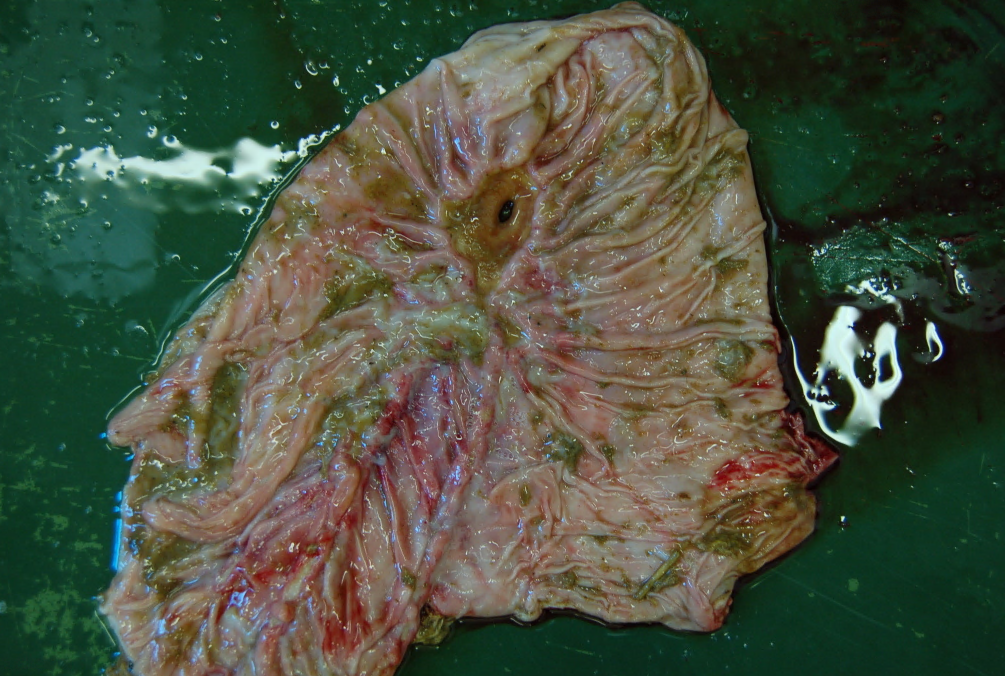
An example of a ruptured abomasal ulcer found in a four-week-old suckler calf
Clostridial disease due to Clostridium septicum was diagnosed in an ewe found in a debilitated state after a cold/wet night. Despite treatment and warming the ewe died. At PM areas of the colon were noted to be dark red in colour and more friable than would be expected. An area of subcutaneous and muscle emphysema located over the right shoulder and right thigh were noted. A sample of the muscle over the right shoulder was sampled and sent for FAT testing. FAT testing did not detect Clostridium chauvoei, Clostridium novyi or Paeniclostridium sordellii but did detect C septicum.
These Clostridial species are responsible for the histotoxic disease presentations. A brief reminder of these below:
- Clostridium chauvoei is responsible for clostridial myositis aka blackleg in cattle which can cause pathology in the skeletal or cardiac muscles.
- Clostridium novyi type B causes infectious necrotising hepatitis aka black disease in cattle and sheep. This disease is usually identified in animals with liver fluke pathology.
- Malignant oedema/gas gangrene is a form of cellulitis and or necrotising myositis due to infection with clostridial species. It is most usually associated with C septicum but other clostridial species have been implicated.
- The most common disease manifestation of Paeniclostridium sordellii is rapidly fatal necrohaemorrhagic and emphysematous abomasitis of lambs aged between four and 10 weeks.
Posted by SRUC Veterinary Services on 19/07/2024